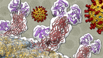
Our research is focused on investigating how different viruses interact with and overcome the complex membranous system that surround and reside within the cell. Using state-of-the-art three dimensional cryogenic electron microscopy techniques we are studding this phenomenon in three main research lines: i) development of nanotransporters/nanocages based on viral capsids, ii) membrane fusion proteins from different viruses including human respiratory syncytial virus (hRSV), human metapneumovirus (hMPV) and coronavirus and iii) rotavirus entry and tropism.
Current projects
Precision therapeutic virus-based nanoparticles
Viral capsids act as self-assembling nanocages, spontaneously forming intricate protein structures that encapsulate and protect a cargo, the viral genome. These nanocages possess the remarkable ability to transport cargo to specific target tissues or locations within the body, delivering payloads to the appropriate subcellular locations with precision and efficiency. This project integrates structural biology, synthetic biology, and recombinant protein production to create advanced protein nanocages. Drawing inspiration from viral-like particles, these nanocages aim to precisely control the distribution of therapeutic cargoes, such as vaccine antigens, drugs, and nucleic acids, while also influencing and directing the immune response.
Rotavirus Host Interactions
Rotaviruses (RVs) represent significant causal agents of infectious gastroenteritis among children under the age of 5, contributing to one-third of all diarrhea-related deaths globally within this demographic. Comprising nine distinct RV groups/species, four of them (A, B, C, and H) have demonstrated infectivity in humans. While RVB, C, and H have been linked to epidemics of severe diarrhea in older children and adults, our understanding predominantly focuses on group A members. Through the integration of advanced structural and molecular virology techniques, this project aims to elucidate the mechanisms enabling these viruses to infect older age groups. By doing so, it seeks to pave the way for the development of novel antiviral strategies against them.
Highlighted publications
- Luque D et al., 2023, 'Equilibrium Dynamics of a Biomolecular Complex Analyzed at Single-amino Acid Resolution by Cryo-electron Microscopy: Equilibrium Dynamics Revealed by cryo-EM Maps', Journal of Molecular Biology, 435, http://dx.doi.org/10.1016/j.jmb.2023.168024, opens in a new window
- Luque D et al., 2022, 'Cryo-EM structures show the mechanistic basis of pan-peptidase inhibition by human α2-macroglobulin', Proceedings of the National Academy of Sciences of the United States of America, 119, http://dx.doi.org/10.1073/pnas.2200102119, opens in a new window
- Luque D and Castón JR, 2020, 'Cryo-electron microscopy for the study of virus assembly', Nature Chemical Biology, 16, pp. 231 - 239, http://dx.doi.org/10.1038/s41589-020-0477-1, opens in a new window
- Jiménez-Zaragoza M et al. 2018, 'Biophysical properties of single rotavirus particles account for the functions of protein shells in a multilayered virus', eLife, 7, http://dx.doi.org/10.7554/eLife.37295, opens in a new window
- Brasch M et al. 2017. Assembling enzymatic cascade pathways inside virus-based nanocages using dual-tasking nucleic acid tags. J Am Chem Soc. 139(4):1512-1519 https://doi.org/10.1021/jacs.6b10948, opens in a new window
Our experts
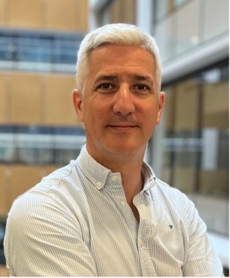
Daniel Luque - Group Leader
Dr Daniel Luque is associate director of the Electron Microscopy Unit and Senior Lecturer at the School of Biomedical Sciences at the University of New South Wales (UNSW). His research is focused on the structural characterisation of viral macromolecular complexes by cryogenic three-dimensional electron microscopy techniques and the development of new applications based on viral capsid nanocages. He holds a Ph.D. degree in Molecular Biology from the Autonoma University of Madrid and was a Postdoctoral Fellow at the Spanish National Centre for Biotechnology. He spent 14 years as Head of the Electron Microscopy Unit of the Spanish Institute of Health Carlos III and group leader at the National Centre of Microbiology. In 2023 he joined UNSW in his current joint positions between the Electron Microscopy Unit the School of Biomedical Sciences.

